The study of plant tissues through fluorescence imaging is becoming increasingly essential in plant biology, biotechnology, and agronomy. To properly investigate these specimens, the use of a slide scanner, originally developed for histology or cytology, proves to be a valuable tool, offering both high-resolution imaging and large field acquisition capabilities.
The Challenge of Imaging Large, Structured Plant Samples
Unlike classical histological samples, plant tissues such as leaves, stems, nodules, and seeds often span several centimeters and display significant morphological complexity. Frequently studied models like Arabidopsis thaliana, tobacco, maize, and others, present anatomical features that require careful spatial context to be properly interpreted.
In many cases, the first step of analysis is to identify and localize areas of interest within large samples. This demands:
A field of view wide enough to encompass entire organs or tissue regions,
Maintaining a sufficient resolution to recognize key structures and guide further analysis.
This balance between resolution and overview is crucial to navigating plant anatomy, which often lacks the consistent and layered structure of animal tissues. A slide scanner capable of capturing large surfaces at multiple resolutions allows researchers to get both the big picture and the cellular detail.
An important consideration when imaging large plant specimens is the time and efficiency required to capture entire tissue sections at high resolution. Traditional imaging approaches, which rely on sequential field-by-field acquisition, often lead to long scan times and require extensive post-processing, especially when dealing with multi-channel fluorescence data. Modern slide scanning systems like InnoQuant equipped with simultaneous multi-channel laser excitation and full width laser scanning acquisition capabilities offer a significant advantage in this context. By capturing multiple fluorescence channels in parallel and eliminating the need for image stitching or reprocessing, these systems dramatically reduce acquisition time while maintaining spatial accuracy and image quality. This efficiency is particularly beneficial for large, complex plant tissues, enabling researchers to image more samples in less time and to generate high-resolution datasets that are immediately ready for analysis.
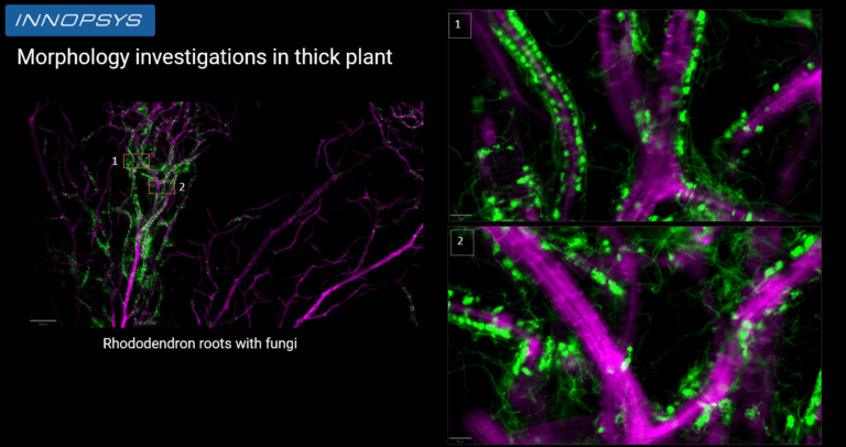
Rhododendron roots in symbiosis with fungi. Full sample captured in 30min at 0,5 µm/px
Autofluorescence and Multi-layered Structures: The Focus Challenge in Plant Biology
One of the most persistent technical challenges in plant fluorescence imaging is achieving optimal focus. Plant tissues are often multilayered, with different types of cellular structures at varying depths. Furthermore, they naturally exhibit strong autofluorescence, which can interfere with the clarity of the signal and complicate focus detection algorithms.
This is where advanced optical systems such as the InnoQuant confocal-like scanner offer a major advantage. By rejecting out-of-focus light, confocal scanning provides a sharp, high-contrast image of the focal plane, even in samples with high background fluorescence. This is particularly useful when scanning leaf surfaces, thick stem cross-sections, or dense root nodules.
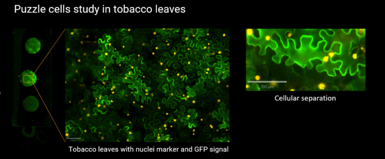
Tobacco leaves with nuclei marker and GFP signal. Acquisition time for the full circle: 13 min.
Throughput and Automation for Comparative Analysis
In addition to imaging performance, slide scanners also bring significant benefits in terms of throughput and automation. The ability to scan multiple samples in a consistent, unattended manner enables the generation of standardized, reproducible datasets. This is particularly valuable when comparing samples across different experimental conditions, genotypes, treatments, or time points.
Automated workflows help reduce variability and support high-content analysis pipelines, making it easier to extract quantitative data from fluorescence signals under controlled acquisition settings.
Conclusion: slide scanner in plant imaging
Using a high-quality slide scanner equipped with confocal capabilities like InnoQuant is a powerful way to overcome the specific imaging constraints of plant specimens.
As plant science continues to expand into imaging-based phenotyping and molecular mapping, slide scanning platforms will play a central role in enabling reproducible, automated fluorescence imaging across a wide range of plant models and experimental conditions.
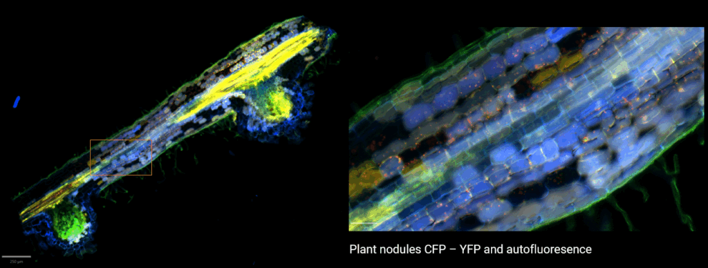
Plant nodule, CFP – YFP and autofluoresence. Acquisition time for the full nodule: 5 min.