Abstract: InnoScan 710-IR microarray scanner, cytokine detection and pGOLD chips
Cytokines are a class of proteins that have emerging roles in diagnostics and therapeutics due to their role in cell signaling and immune system response1,2. Many key cytokines including interleukin-6 (IL-6) and interleukin-1 beta (IL-1b), can range in concentration in the serum form femtomolar (fM) to nanomolar (nM) levels3,4. A 6 log dynamic range of cytokines maykes it difficult for conventional ELISA technology to accurately assess because of a 2-4 log dynamic range limit5. The InnoScan 710- IR scanner from Innopsys, when coupled with pGOLD slides from Nirmidas Biotech, is able to quantify IL-1b levels over the relevant 6 logs of dynamic rage thanks to the near-infrared detection of the InnoScan 710-IR scanner and the metal- enhanced fluorescence (MEF) capabilities of the pGOLD slides.
Introduction
Cytokines play a crucial role in cell signaling and are a key indicator of the state of the body’s immune system. Cytokine detection can aid in diagnosis of a range of diseases including cardiovascular disease6 and respiratory issues7. However, cytokines are small proteins (5-20 kDa in size) that are expressed across a wide concentration range, from sub-pg/ml8 up to hundreds of ng/ml3. Currently used, widely available assay technologies, including ELISA and bead-based technology, have detection limit of ~1 pg/ml, thus missing cytokine that may be present at low concentration. Likewise, most ELISA and other assay technology tend to scan fluorescence in the visible region, which present high endogenous auto- fluorescence for biological samples, limiting the sensitivity. This lack of reliable quantification of cytokines in serum and plasma may be hindering their use as diagnostics makers. The short-comings of the current assay techniques are two-fold; first, the instrumentation for detection lacks the necessary wavelength range for low background, and secondly, the signal for detection is not present at high enough levels for lower concentration biomarker.
Innopsys has created a microarray scanner that can directly address the issue of range by offering the extended range (XDR) option in its InnoScan 710-IR microarray scanner. For low concentration samples such as biologically relevant levels of cytokines, InnoScan 710-IR is capable of scanning fluorescence in the near-infrared (NIR) region with excitation wavelengths 670 and 785 nm.
In this region, endogenous auto-fluorescence virtually drops to zero, greatly improving the lower limit of detection. Also important, the number of NIR fluorescent dyes with high quantum yield that are capable of bioconjugation has increase in recent years as well. The InnoScan 710-IR microarray scanner can be also used with the XDR mode. Using XDR mode, the signal dynamic range is extended from 4 logs to a 6 logs. This allows retrieval of all the biological information in complex samples at higher concentrations. The XDR function has been shown to capture more exploitable data without any modification of its distribution, which is crucial when comparing samples with different expression levels.
Similarly, Nirmidas Biotech, Inc., has recently commercialized a plasmonic chip technology, called pGOLD, wich amplifies fluorescent signal up to 100- fold, allowing for detection of biomarkers at much lower concentrations than previously achievable8,10-13
pGold chips use the same protocols and have the same shape as standard glass microscope slides and amplify fluorescence signal maximally in the red and NIR rage of fluorescence. In this application note, the NIR scanning capabilities of the InnoScan 710-IR are combined with the signal-amplifying effect provided by pGOLD chips in order to detect sub-pg/ml concentrations of IL-1b.
experimental section
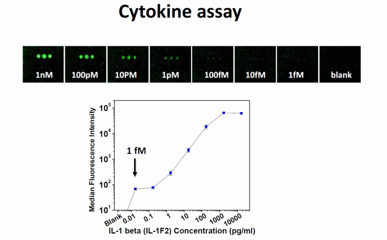
A functionalized pGOLD slide was divided into 8 sectors, three spots were printed with anti-IL-1b monoclonal antibody, acting as the capture antibody, using albumin (BSA) in phosphate buffer saline (PBS). Afterwards, each section of the pGOLD slide was incubated with a different concentration of human IL- 1b cytokine in a BSA/PBS solution, ranging from 1 fM up to 1nM along with one blank section. After washing, each section was incubated with 5 nM of anti-IL-1B-IR800 polyclonal antibody. After incubation with the detection antibody-fluorophore conjugate, washing removed free, unbound antibody and the pGOLD slide was ready for scanning.
The InnoScan 710-IR instrument was used for scanning with an excitation wavelength of 785nm. The PMT gain was set to 20 while the pixel size for scanning was set to 20 µm due to the large spot size; the InnoScan 710-IR is able to achieve resolution as low as 3 µm pixel size.
results and discussion: InnoScan 710-IR microarray Scanner excels at cytokine detection in conjunction with pGOLD chips
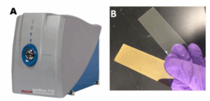
pGOLD slides rely on using metal-enhanced fluorescence (MEF) created by the gold nano-island surface structure to amplify fluorescence
signal in protein sandwich assays. MEF as a phenomena has been known to exist for several decades but just recently optimized by research at Stanford University8,10-13, which Nirmidas Biotech has commercialized as its pGOLD slides (Figure 1b). pGold slides, with the same morphology as typical glass slides, have a nano-structured gold coating that can increase the fluorescence of traditional reporter dyes,
requiring a high-performance microarray scanner capable of extending out into the NIR range. Figure 2 shows data from the assay of human IL-1b cytokine on a pGOLD slide, achieving detection down to 1 fM of the cytokine. As demonstrated here, the InnoScan 710- IR is able to easily detect the fluorescenceemission of IR800 while offering the optimum wavelength region to detect biomarkers such as cytokine IL-1b below 0.1 pg/ml concentration with a >5 log dynamic range (Figure2).
references
- O’Shea, John , Massimo Gadina, and Robert D. Schreiber. Cell
109.2 (2002): S121-S131.
- Dantzer, Robert, et Nature Reviews Neuroscience 9.1 (2008): 46-56.
- Damas, Pierre, et Annals of surgery 215.4 (1992): 356.
- Moldoveanu, Andrei , Roy J. Shephard, and Pang N. Shek.
Journal of Applied Physiology 89.4 (2000): 1499-1504.
- Martins, Thomas , et al. American journal of clinical pathology 118.3 (2002): 346-353.
- Mendall, A., et al. Heart 78.3 (1997): 273-277.
7. Bonfield, Tracey L., et al. American journal of respiratory and critical care medicine 6 (1995): 2111-2118.
8. Zhang, Bo, et Chem. Sci. (2014).
9. Füzéry, Anna , et al. Clinical proteomics 10.1 (2013): 13.
10. Tabakman, Scott M., et al. Nature communications 2 (2011):
11. Zhang, Bo, et Nano Research 6.2 (2013): 113-120.
12. Zhang, Bo, et PloS one 8.7 (2013): e71043.
13. Zhang, Bo, et Nature medicine 20.8 (2014): 948-953.