
Line-scanning microscopy: a different approach to fluorescence slide imaging
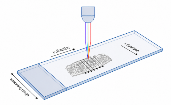
Line-scanning microscopy is an imaging approach in which fluorescence signals are acquired continuously, line by line, as the optical axis scans across the sample. At Innopsys, this principle is used in both the InnoScan and InnoQuant platforms to generate high-resolution fluorescence images over large areas, up to entire microscope slides.
From fluorescence signals to digital images
Unlike conventional field-of-view imaging systems, which capture successive rectangular images using camera-based detectors, line-scanning systems build the image progressively during the scan.
In InnoScan and InnoQuant instruments, fluorescence is excited by lasers and detected using photomultiplier tubes (PMTs). The signal collected along each scanned line is converted into individual pixels, which are progressively assembled to reconstruct the complete digital image.
Why does this matter?
Camera-based whole-slide imaging typically relies on the acquisition of many individual fields of view. These fields must then be combined to reconstruct the complete image, a process known as stitching.
In addition, illumination and detector response are not always perfectly uniform across a camera field. Image-processing algorithms such as shading or flat-field correction are therefore commonly used to compensate for these variations.
Full-width line scanning fundamentally changes this imaging workflow.
Because the signal is acquired through a highly consistent optical path throughout the scan, Innopsys systems can achieve field uniformity of up to 98%. The resulting image does not require conventional field stitching or shading correction.
A key advantage for fluorescence quantification
These characteristics are particularly important when fluorescence intensity itself carries quantitative information.
Variations in illumination, detector sensitivity or image correction between different areas of a slide can introduce biases when comparing fluorescence intensities. Avoiding these sources of variability provides a major advantage for applications in which signal intensity must be measured reliably.
This principle has historically been particularly valuable in microarray analysis, where fluorescence intensity directly reflects the abundance of a biological target.
The same requirement increasingly applies to quantitative immunofluorescence. Whether comparing biomarker expression between different regions of a tissue, between slides, or across experimental batches, a homogeneous and reproducible imaging chain helps ensure that measured differences reflect the biology of the sample rather than variations introduced by the imaging system.
From microarrays to quantitative fluorescence imaging
Line-scanning technology therefore provides more than a way to digitize large fluorescent samples.
By combining laser excitation, PMT detection and continuous image formation, it creates a highly uniform imaging environment particularly well suited to quantitative fluorescence measurements.
With InnoScan for microarray imaging and InnoQuant for fluorescence whole-slide imaging, Innopsys applies the same fundamental principle to applications ranging from molecular assays to quantitative tissue imaging.