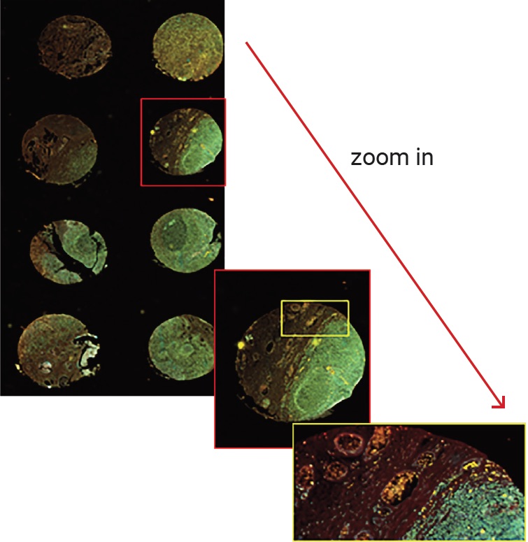
Protein detection by fluorescence-labelled antibodies on tissue sections organzied in tissue microarrays is a widely used technique for the biomarker screening in complex diseases such as cancer to define biomarker expression on tumor cells and its distribution on a defined tissue.
Contrary to microscopes in which the image is built of the stitching of different field of views, the Innopsys’ InnoScan 1100 microarray scanner builds the images form a real and accurate fluorescence PMT detection at a very high resolution without any stitching or tiling process. This ensure the best image quality for tissue microarrays.
The scan of the whole tissue microarray slide is done under the same laser power and PMT gain detection generating highly reproducible results and allowing to compare between different samples.Innopsys’ in content focusing algorithm allows to get precise focusing all along the scan without user intervention.