
Digital pathology (DP), also known as virtual microscopy or whole slide imaging (WSI), is an alternative to conventional microscopy. Beyond enabling a fully digital workflow for anatomical pathology departments, this technology consists of digitizing a glass slide to produce a digital image. This technology therefore meets the needs of microscope users who seek to visualize their slides, image them, interpret them and easily share their images. We therefore prefer to use the terminology of whole slide imaging to not restrict this technology to the characterization of pathological tissues.
What are the limitations of conventional microscopy?
The invention of the microscope by Zacharias Janssen at the end of the 16th century revolutionized Science and particularly Biology by allowing, through the combination of an objective and an eyepiece, to magnify the image of a small object and to separate the details of this image that were not observable by the human eye. Microscopy thus allows access to the invisible.
However, even though microscopy has evolved over the centuries, the user still faces some technical obstacles. The field of view of an objective lens is often smaller than the sample size. Moreover only one person controls the microscope, this one imposes his choice of view to the other. Thus, a series of acquisitions is necessary to image the whole sample. All the captured images are then aligned and stitched into larger mosaics to reconstruct the whole slide. This process is really time consuming and can generate image reconstruction artifacts.
Why is whole slide imaging changing the labs workflow?
In conventional microscopy, we place the glass slide under the microscope and visualized it. There is therefore a viewer in one place at a time. We keep the slide to re-imaged it if necessary. However, during storage, there is a risk of deterioration or breakage. This process is valid for all samples generated by the laboratory. Thus, “physical” slides accumulate and storage choices have to be made. Furthermore, re-imaging these slides by a different user lead to a different view from the previous time and raise questions.
Labs using whole slide imaging acquire a high resolution digital image of the whole glass slide with a scanning device. The digital file obtained allows to visualize the slide as under the microscope however, this virtual transformation allows to store the image on a server. We can then visualize the virtual slide several time without damaging it. People despite their geographical location can share and analyze it. Different dedicated software are available to visualize and/or analyze these digital slides (open source or not). Moreover, scanners are more and more automated. A user will be able to image successively several slides in a minimum of time and with the least possible intervention.
Why associate WSI and Innopsys?
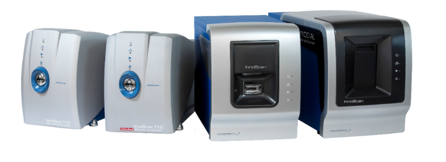
Innopsys is a pioneer in whole slide imaging for 20 years. Our business core is the manufacturing of microarray scanners, the Innoscan scanners. Our scanners allow to digitize microarrays glass slide into high resolution microarrays virtual slide. We continue to improve these scanners by combining our know-how in mechanics, optics, embedded systems and IT to optimize the image acquisition for fast, high-resolution scanning with the least possible human intervention. We are currently using these skills to develop our new scanner, InnoQuant (coming soon), a high-resolution whole slide scanner for tissue and cell sections.
How to proceed to perform whole slide imaging with Innopsys scanners?
First, insert the slide (or several) in the scanner which is a closed enclosure. Secondly make an entire slide preview at a very low resolution to define the area of interest. Then adjust the parameters of sample illumination and the signal detection settings. To be compatible with as many samples as possible, it is possible to choose a focus methodology to compensate the heterogeneous thickness of the sample and obtain a mapping of the acquisition position. Finally, scan at the desired resolution line by line, from one corner of the area to the opposite corner to obtain a digital image of the glass slide. #WSI by Innopsys.
Thus, whole slide imaging improves conventional microscopy and facilitates laboratory workflow. Over the last few years, this technology has grown significantly in various fields. As indicated by the other name of WSI, Digital pathology, pathologists are also adopting this technology. The College of American Pathologists has established guidelines for clinical validation. The FDA has also approved the use of this technology for primary diagnosis since 2017. In addition, the COVID-19 pandemic has exacerbated the presence of the virtual in our daily lives and the WSI also finds all its advantages in research, clinical or education initiatives.
Exemple of InnoQuant workflow
